
Development of Color-X-ray Histology
ID# 2014-4240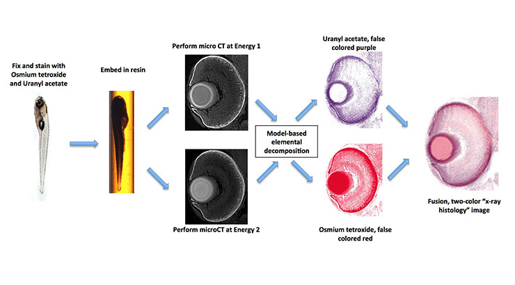
Technology Summary
This invention allows for the non-invasive, rapid generation of three-dimensional (3D), multi-colored images of tissues and optically opaque organisms (e.g. zebrafish) through the use of two or more heavy-metal X-ray dyes.The major advantage of this invention is that it allows for visualization of two or more X-ray dyes in the same tissue. Previous methods cannot differentiate between signals from multiple X-ray dyes in the same tissue, andare labor-intensive and imprecise. This method is fast, requires no cutting of tissue sections, and allows for the generation of high-resolution (1-2 µm), multi-colored 3D reconstructions. Use of multiple stains allows for identification of specific cell types within the sample. Rapid identification of cells can be used to phenotype whole opaque organisms.
Application & Market Utility
This invention may be of particular interest to companies focused on optics and imaging, as well as to developers of diagnostic products based on novel imaging technologies. Because this invention can differentiate between healthy cells and diseased cells, this invention may also be of interest to pathologists.
Next Steps
Seeking licensing partner for commercialization.