
MRI-Active Biodegradable Polymers
ID# 2016-4527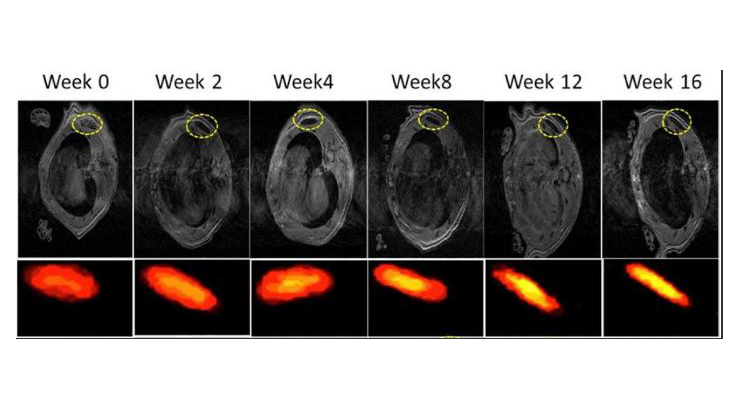
Technology Summary
Although it is well known in the tissue engineering field that an implantable scaffold’s degradation rate should match the tissue regeneration rate, lack of a quantitative method for in vivo validation has limited progress in this area. To address this issue, researchers at Penn State have created a family of citrate-based biodegradable polymers that comprise a magnetic resonance imaging (MRI) contrast agent either integrated into or pendent from their polymeric backbones. These materials can also incorporate photoluminescent moieties, providing dual-imaging capabilities to track tissue regeneration and scaffold degradation processes non-invasively. MRI-active, NIR-excitable implanted biodegradable polymers permit 3D imaging of structure and volume changes, even in deep tissue. The figure above shows T1-weighted images of scaffolds implanted within deep tissue at different time points.
Application & Market Utility
This technology enables an unprecedented understanding of scaffold degradation in situ that is vital to the biomaterial design process for regenerative engineering. The ability to effectively image biodegradable scaffolds implanted within deep tissue (i.e., depths > 2 cm) and monitor degradation over time has major implications for biomaterial designers. It is now possible to design a scaffold with a tunable degradation rate, implant the scaffold within deep tissue, and non-invasively validate its degradation profile in situ and in vivo.
Next Steps
Seeking research collaboration and licensing opportunities.