
Spectrally Encoded Imaging Using Band-Shifting Fluorophores
ID# 2018-4803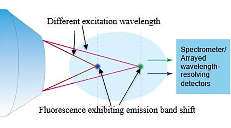
Technology Summary
Penn State researchers have developed a novel concept of parallel axial imaging using band-shifting fluorescent probes. The excitation wavelengths are first mapped to different spatial locations in the axial direction by using chromatically aberrated excitation light. The wavelength-space encoding is then transferred to a fluorescence signal through the use of band-shifting fluorescent probes, which are fluorophores that exhibit an excitation-dependent emission band (traditional fluorophores produce the same emission band regardless of the excitation wavelength). This concept is similar to using multiple wavelength channels to carry digital bits of information in parallel in an optical fiber (i.e., wavelength division multiplexing), and can enable faster single-photon or multi-photon fluorescence imaging by eliminating or reducing time-consuming axial scanning.
Application & Market Utility
The present technology addresses a limitation of laser scanning microscopy, particularly epi-fluorescence confocal microscopy and multi-photon fluorescence microscopy, which is slow axial scanning speed. It is believed that this technology can improve axial scanning speed by 10x. Accordingly, parallel axial imaging could expand the capability of two-photon imaging for real-time monitoring of fast biological processes at multiple depths using a micro-endoscopic imaging head.
Next Steps
Seeking research collaboration and licensing opportunities.