
Transparent Ultrasound Transducers for Photoacoustic Imaging
ID# 2018-4838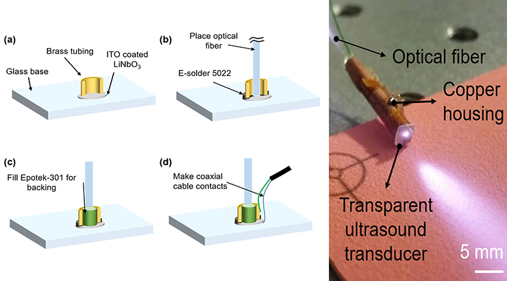
Technology Summary
Photoacoustic imaging (PAI) has emerged as one of the most promising biomedical imaging modalities as it maps optical contrast of deep tissue with high spatial resolution. PAI uses pulsed-light excitation of the tissue to cause wideband ultrasound generation due to transient thermoelastic expansion of light-absorbing molecules such as melanin and hemoglobin. In current PAI systems, light is delivered to the tissue by bending the beam around the transducer or through an opening in the transducer. This leads to bulky devices, limits possible device architectures, and constrains PAI efficacy in terms of depth, resolution and speed. Penn State researchers have solved these issues by incorporating a transparent transducer material (e.g., lithium niobate). The resulting device designs are low-cost, easily manufacturable, and compatible with conventional clinical ultrasound electronics.
Application & Market Utility
PAI is ideal for imaging in areas where differences in optical absorption exist. These contrast differences can occur naturally, such as in a large lipid pool or thin fibrous cap in vulnerable atherosclerotic plaques or angiogenesis in tumors – an early indicator of various cancers. Contrast can also come from exogenous sources, such as contrast-agent-labeled antibodies. Permitting the light and sound to share the same beam path will enable miniaturization of PAI devices, allowing PAI to finally be utilized in space-constrained applications like endoscopic imaging.
Next Steps
Seeking research collaboration and licensing opportunities.